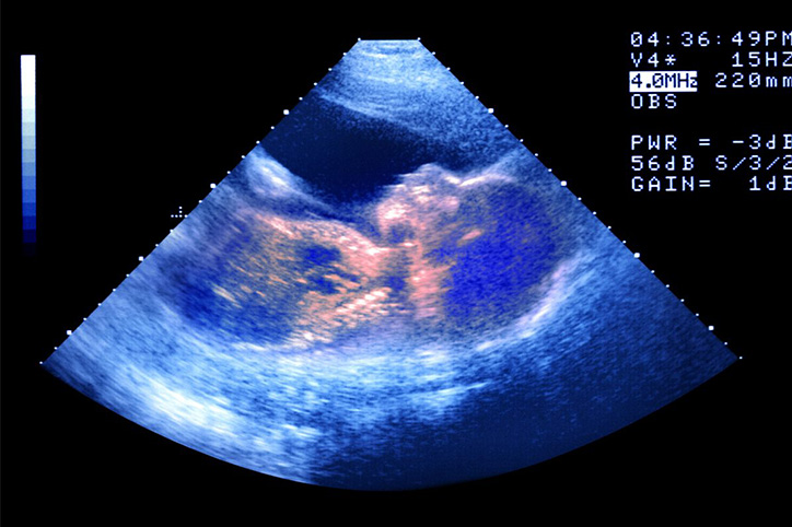
Given that I travel so frequently between Tel Aviv and New York City, I had doctors in both cities during my pregnancy journey and the differences were shocking! For one, in America you typically get almost no ultrasounds, whereas in Israel you are seeing your doctor for one every four weeks – or more if there’s an issue. I had hyperemesis gravidarum, so in those first 16 weeks until my constant nausea subsided there were times I was getting more than one ultrasound a week.
“In most pregnancies women only get two ultrasounds: a dating ultrasound around eight weeks and then an anatomy scan around 18-20 weeks,” says Megan Pallister, MD, FACOG. At the first ultrasound the fetus is about 2 cm long or roughly the size of a raspberry. All major organs are forming and you can hear the heartbeat by now. Blood cells are also developing. The neural tube has closed (it does this by week six), so this is why you need your folic acid intake to start before you even start trying to attempt pregnancy. The neural tube is what the brain and spinal cord will form from.”
Then, Dr. Pallister explains, the anatomy scan measures the anatomy of the baby (heart, kidneys, brain, etc), the size and growth of the baby, information about the placenta and measures the amniotic fluid. “After 20 weeks, we measure the fundal height at every visit— at 20 weeks, the top of the uterus is usually right at the level of your belly button and every week it grows about one centimeter. If our measurements are ever off, we can get an ultrasound to see how the baby is growing.” Another common reason to get follow up ultrasounds after the anatomy scan is if the doctor was not able to evaluate all of the fetal anatomy on the first scan. “There are a list of structures we need to see in detail to rule out anomalies,” says Dr. Pallister. “Some of these include the spine, kidneys, brain and heart. Often babies may be facing the wrong way for us to get a clear picture of these structures so we will do a follow up ultrasound a few weeks later.”
In Israel, at the very minimum women get what is called a “nuchal translucency scan” (which tells parents if their baby is likely to have a chromosomal abnormality) and “early anatomy scan” (a check of the baby to see if all the major structures are developing normally) in addition to the aforementioned two scans. Both scans take place in the weeks between the dating ultrasound and the anatomy scan. I was told that some problems can be seen before 18 weeks and it is beneficial to know if there is an issue sooner rather than later. While this is standard in Israel, it is not in America, but something you can request. Personally, the more scans I got the more re-assured I felt and there is no harm to Mom or baby when doing these ultrasounds.
Dr. Pallister noted that in certain cases, early anatomy scans can be helpful. “In patients with a history of fetal anomalies or at risk for a genetic syndrome, a nuchal translucency around 11-13 weeks can be done.”
It’s a personal choice, but knowledge is power and just because you don’t have a reason to suspect that there is an issue, it doesn’t mean you shouldn’t consider requesting additional ultrasounds if that may help with peace of mind.


